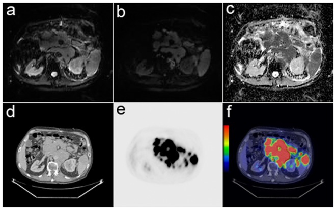
Figure 1. Diffusion-weighted MRI and PET/CT images showing the abdominal region tumor in a 76- year old male patient with diffuse large B-cell lymphoma.

(a) B0 image. (b) Diffusion-weighted image with b value 800 s/mm2 showed the hyperintensity tumor, but it was not able to depict diffuse spleen involvement. (c) The corresponding ADC map showed the hypointensity tumor with ADCmin 0.34×10⁻³ mm2/s and ADCmean 0.68×10⁻³ mm2/s. (d) Axial CT image. (e) FDG-PET image. (f) The fused PET/CT image showed the active tumor and spleen involvement with SUVmax 23.9.
