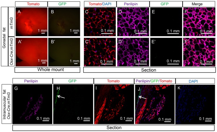
Figure 3. Osx-Cre does not mark non-bone marrow adipocytes.
(A–B) Images for direct fluorescence from tdTomato (A) or EGFP (B) in whole-mount gonadal fat depots from two-month-old Osx-Cre; R26-mT/mG mice. (C–F) Direct fluorescence for tdTomato (C) and immunofluorescence for perilipin (D) and EGFP (E) on sections of gonadal fat depots from two-month-old Osx-Cre;R26-mT/mG mice. (G–K) Imaging of longitudinal sections of an intramuscular fat depot associated with a tibia from two-month-old Osx-Cre;R26-mT/mG mice. G: perilipin immunofluorescence; H: EGFP immunofluorescence; I: direct fluorescence for tdTomato; J: merged view of G–I; K: DAPI staining. Arrow: GFP-positive periosteum.

