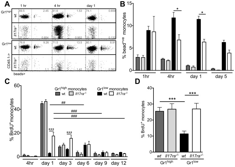
Figure 7. Kinetics of labeled Gr1high and Gr1low monocytes in the presence and absence of Il17ra-/-.
(A,B) Monocytes in mixed chimeric wt/Il17ra-/- mice were labeled with fluorescent latex beads (A). The proportion of labeled CD11b+CD115+ cells among each monocyte subgroup (Gr1high and Gr1low monocytes) within each genotype was assessed over time (B, n = 4-5, 2 independent transplantations, Bonferroni after ANOVA). (C) Dividing cells in mixed bone marrow chimeric wt/Il17ra-/- mice labeled with a single BrdU injection and peripheral blood monocytes were analyzed for BrdU incorporation at the indicated timepoints. In every animal, the proportion of labeled cells among Gr1high and Gr1low monocytes of either genotype was assessed over time (n = 4, * indicates sign. differences between wt and Il17ra-/- Gr1low monocytes at the indicated timepoints, # indicates a sign. change over time within wt (black bars) or Il17ra-/- (white bars) Gr1low monocytes, analysis with Bonferroni after ANOVA). (D) Proliferation of bone marrow Gr1high and Gr1low monocytes of both genotypes was assessed 20 h after BrdU injection (n = 7 from 2 independent transplantations, Bonferroni after ANOVA).

