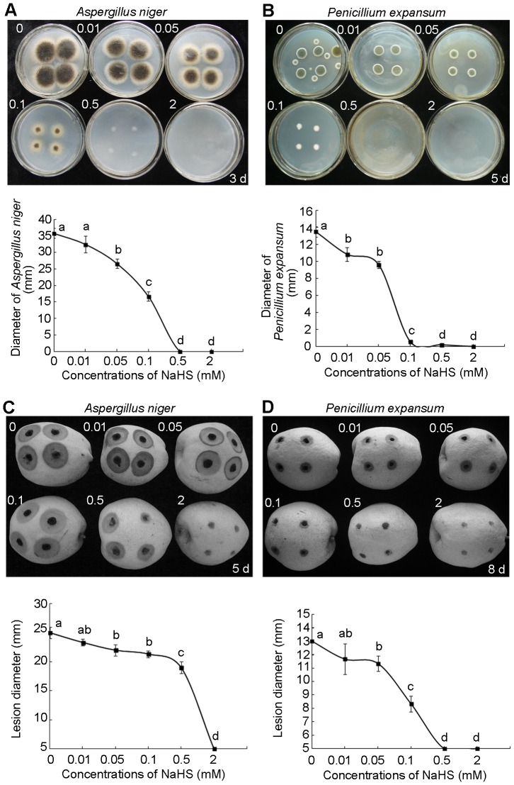
Figure 5. H2S inhibits the growth of Aspergillus niger and Penicillium expansum.
A. niger (A) and P. expansum (B), isolated from pear slices, were cultured on medium and subjected to the fumigation of different concentrations of NaHS solutions for 3 and 5 days respectively. The upper photographs of (A) and (B) indicate the growth of fungi subjected to different concentrations from left to right, upper to lower 0, 0.01, 0.05, 0.1, 0.5 and 2 mM NaHS, and the lower part of figure shows the diameters of fungi clones. The upper photographs of (C) and (D) show the pears infected by A. niger and P. expansum for 5 days and 8 days, respectively. The pears were subjected to different concentrations from left to right, upper to lower 0, 0.01, 0.05, 0.1, 0.5 and 2 mM NaHS, and the lower part of (C) and (D) shows the diameters of wounds caused by fungi. Data are presented as means ± SD (n = 4). Different letters indicate significant differences (p<0.05) between the treatments.

