
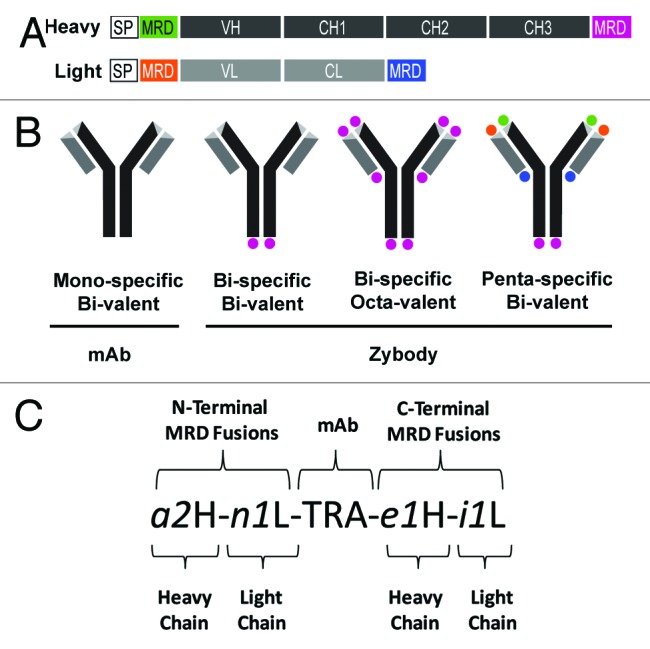
Figure 1. Schematic diagram of zybody assembly and nomenclature. (A) Schematic representation of immunoglobulin heavy (H) and light (L) chain genetic fusions that constitute a penta-specific zybody with four unique MRDs. Mouse γ and κ chain signal peptides (SP) were fused to the heavy and light chains, respectively. Signal peptides, Ig variable (V) and constant (C) domains, and MRDs are encoded in one contiguous open reading frame. (B) A schematic representation of a mono-specific antibody scaffold along with three exemplary zybodies, illustrating varying degrees of specificity and valency. (C) The zybody nomenclature identifies the antibody scaffold by a capitalized three-letter code (e.g., TRA for trastuzumab, ADA for adalimumab, CET for cetuximab, PAL for palivizumab). MRDs are indentified with an italicized, lowercase alphanumeric code (e.g., a2). The letter of the MRD code indicates the MRD target (e.g., i for IGF-1R, e for EGFR, n for integrin αvβ3, a for Ang2, c for ErbB3), followed by a unique numeric identifier and either an H (heavy) or an L (light), indicating the chain to which it is fused. The terminus to which the MRD is fused can be inferred from the position of the MRD identifier relative to the scaffold.