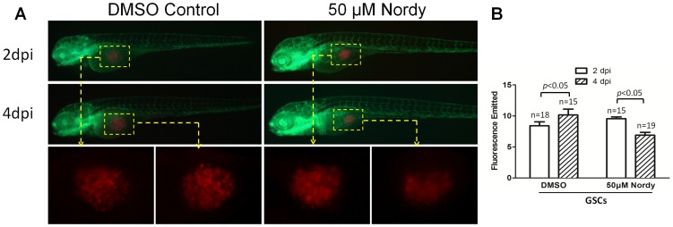
Figure 5. Effects of Nordy on the proliferation of GSCs within zebrafish embryos.
A. Representative merged images of GSC proliferation with/without Nordy treatment within zebrafish embryos. B. Quantitative analysis of the emitted fluorescence of RFP-labeled GSCs with/without Nordy treatment at 2 dpi and 4 dpi. At higher magnification, the images showed the injected the RFP labeled GSCs accumulate within the embryos (yellow broken box). Red: injected U87-RFP cells; green: GFP fluorescence of angiogenesis in Tg (fli1:EGFP)y1 embryos.

