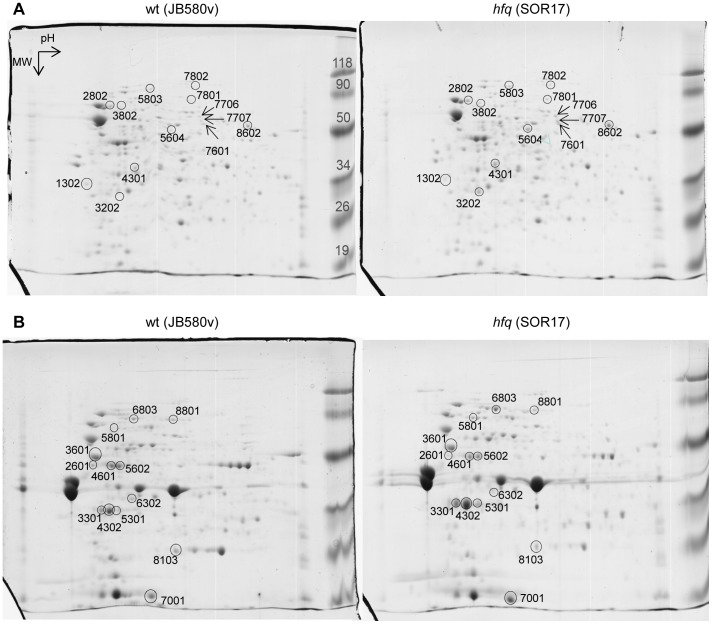
Figure 2. 2-DE analysis of total soluble (A) and total membrane (B) proteins stained with Coomassie blue.
Bacteria were grown in triplicate at 37°C for 5 h. One representative gel per strain is shown. Proteins were separated in 2-DE gels (for all gels: pH range 3–10, molecular weight (MW) range 15–150 kDa). Highlighted spots were identified by mass spectrometry (see Table 3). MW marker size is indicated in kDa.

