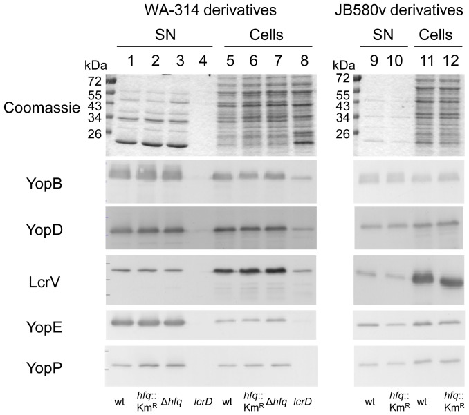
Figure 8. Analysis of Yop proteins secreted by Y. enterocolitica.
Proteins secreted into the supernatant (SN, lanes 1-4, 9–10) and proteins from total bacterial cell extracts (Cells, lanes 5–8, 11–12) were analyzed by Coomassie blue staining (upper panel) and by immunoblotting using antibodies specific for YopB, YopD, LcrV, YopE and YopP. Loading was as follows: molecular weight markers (in kDa); 1 and 5, parental strain WA-314; 2 and 6, hfq mutant SOR3; 3 and 7, hfq mutant SOR4; 4 and 8, TTSS-defective lcrD mutant strain WA-314(pYV-515); 9 and 11, parental strain JB580v; 10 and 12, hfq-negative strain SOR17.

