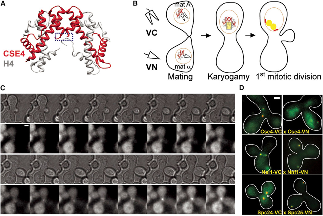
Figure 1. More Than One Cse4 Molecule Is Deposited at the Centromere in S Phase.
(A) Model of the H4-Cse4 heterotetramer shows that the carboxyl termini of the two Cse4 molecules (blue box) are separated by more than 2 nm.
(B) Schematic of approach to monitor the deposition of two or more Cse4 molecules at the centromere. Mating of two haploid strains expressing Cse4-VC (carboxyl fragment of Venus) and Cse4-VN (amino fragment of Venus) is followed by karyogamy, after which the newly diploid cell enters its first mitotic division. The two Cse4-BiFC species can interact with each other only after cell fusion. Therefore, development of Venus fluorescence in the zygote relative to the mitotic spindle (demarcated by red spindle pole bodies) will indicate Cse4 deposition at the centromere.
(C) Time-lapse microscopy showing the development of a single Venus fluorescence focus within the zygote after bud emergence, which coincides with entry into S phase (top panels, transmitted light images; bottom panels, maximum projection YFP images acquired every 5 min, scale bar represents ~ 2 µm).
(D) Colocalization of the BiFC fluorescence with a spindle-pole body marker (either Tub4-mCherry or Spc97-mCherry) confirms that the fluorescence is associated with centromere or kinetochore clusters (scale bar ~ 0.96 µm).

