FIG. 2.
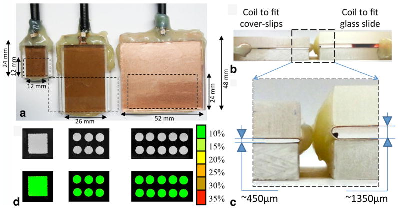
A set of five histological slide coils were developed to accommodate both different sample sizes and slide setup. a: The coils were designed in three sizes to house encasing based on dual coverslips for tissue sections up to 100 μm thick (insert H ≈ 450 μm). The smallest coil (left: W = IL = 12 mm, OL = 24 mm) was designed to insert 24 × 12 mm coverslips. The middle coil shown is of similar size (W = 26 mm, IL = 24 mm, OL = 48 mm) to the structure previously reported (35) to enable the partial examination of standard size slides (50 × 24 mm). The larger structure (W = 52 mm, IL = 24 mm, OL = 48 mm) shown at right permits the full coverage of slides of the same size. b: Another two coils with identical size to the two largest probes were additionally designed with a greater height (H ≈ 1350 μm instead of H ≈ 450 μm) to fit the coverslip/glass slide set up. c: All the coils proved to have an excellent RF planar homogeneity assessed experimentally using MRI with a phantom composed by a thin layer of water doped with 2.5 mM Gd-DTPA sandwiched between two coverslips. As indicated in the color scale, the green level represents less than 10% deviation from the average signal intensity measured by the ROI outlined in the map (white dashed line).

