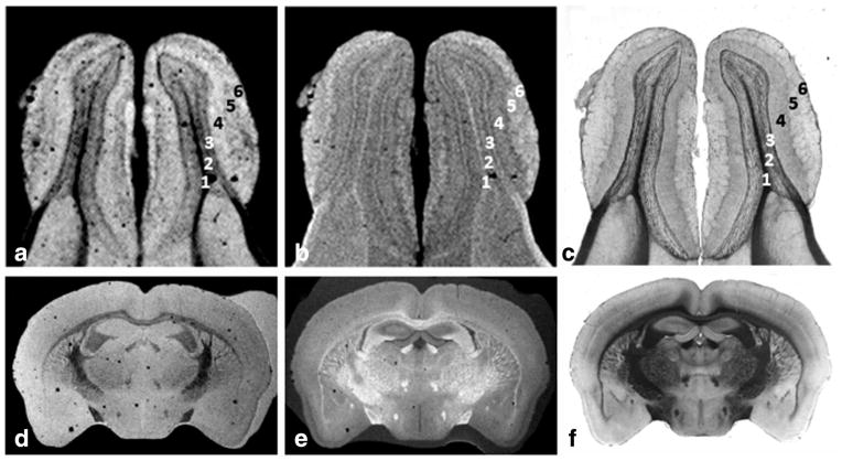
FIG. 6.
The highly detailed image examples shown were obtained from 60 μm tissue sections using the smallest and most sensitive coverslip histology coil designed in this study (W = 12 mm, IL = 12 mm, OL = 24 mm, H = 450 μm). The first image set (a) and (b) correspond respectively to T2*- and T1-weighted images with 30 μm in-plane resolution obtained from the mouse olfactory bulb coregistered with the corresponding light microscopy (c). The MRI contrast on both images helps identify the following cell layers: (1) olfactory ventricle, (2) combines the internal plexiform layer, granule cell layer and ependymal layer, (3) mitral cell layer, (4) external plexiform layer, (5) glomerular layer, (6) olfactory nerve layer. The coil can also accommodate coronal mouse brain sections depicted by the example of a 50-μm in-plane MRI showing clearly the white matter track and different small tissue structures in (d) T2*- and (e) T1-weighted images in perfect alignment with (f) histology.

