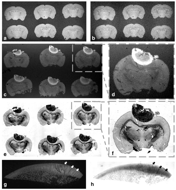
FIG. 7.
Both large coverslip and glass-slide coils can be used for high throughput acquisitions by scanning simultaneously multiple tissue sections (a–f). This setup can also accommodate large size samples (g–h). The difference in SNR between T2*-weighted brain images acquired in (a) SNR = 47 and (b) SNR = 28 illustrates the 3× greater filling factor of the dual coverslip coil (W = 52 mm, IL = 24 mm, OL = 48 mm, H = 450 μm) compared with the identical structure but capable of housing samples embedded in traditional coverslip/glass slide setup that are much thicker (coil with equal dimensions but H = 1350 μm). The example shown in (c) depicts the T1 brightening of a melanoma tumor in a set of 6 × 40 μm mouse brain sections premounted in a glass slide and acquired with a 60-μm in-plane resolution where (d) corresponds to a magnification of the boxed area in (c) and (e and f) the matched histology. The image example in (g) corresponds to a 100-μm in-plane gradient echo image of a 5-μm human trochlear cartilage (SNR = 33 for an 8 h scanning time) and (h) histology of the same sample using Alcian blue dye to stain glycosaminoglycans and help identify the loss of in cartilage integrity highlighted by the contrast from both imaging modality depicted by the arrows in g and h.

