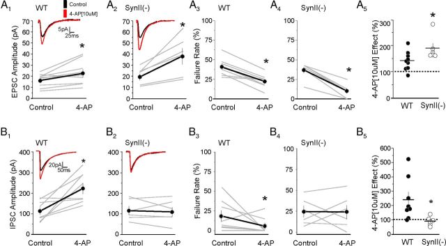
Figure 3.
4-AP promotes excitatory and inhibitory transmission in a balanced way at WT synapses but creates an imbalance toward excitation at SynII(−) synapses. A, EPSCs recorded in slices derived from young presymptomatic SynII(−) mice exhibit an enhanced sensitivity to 4-AP (10 μm). A1, Effect of 4-AP on EPSC amplitude in WT slices (p = 0.014, n = 9). A2, Effect of 4-AP on EPSC amplitude on SynII(−) slices (p = 0.011, n = 6). A3, Effect of 4-AP on the EPSC failure rate in WT slices (p = 0.0002, n = 9). A4, Effect of 4-AP on the EPSC failure rate in SynII(−) slices (p = 0.0001, n = 6). A5, The synaptic facilitation induced by 4-AP effect is significantly (p = 0.049) stronger at SynII(−) synapses. The scatter graph represents the ratio of EPSC amplitudes with and without 4-AP. Data collected from 4 WT mice and 3 SynII(−) mice. A1, A2 insets, Average EPSCs (50 sweeps) recorded from representative experiments. Thin gray lines (A1–A4) and circles (A5) represent individual experiments. The depicted values correspond to the average of 50 EPSCs. Asterisks indicate a statistically significant difference. One-sided paired t test was used to determine an increase in synaptic response upon 4-AP application for the experiments depicted in A1–A4. Two-sided unpaired t test was used to compare the results depicted in A5 to determine the difference between genotypes. B, IPSCs recorded in slices derived from young presymptomatic SynII(−) mice exhibit diminished sensitivity to 4-AP (10 μm). B1, Effect of 4-AP on the IPSC amplitude recorded in WT slices (p = 0.010, n = 8). B2, Effect of 4-AP on the IPSC amplitude recorded in SynII(−) slices (p = 0.67, n = 6). B3, Effect of 4-AP on the IPSC failure rate recorded in WT slices (p = 0.05, n = 8). B4, Effect of 4-AP on the IPSC failure rate recorded in SynII(−) slices (p = 0.44, n = 6). B5, The effect of 4-AP is significantly (p = 0.036) stronger at WT synapses. Data collected from 4 WT mice and 3 SynII(−) mice.

