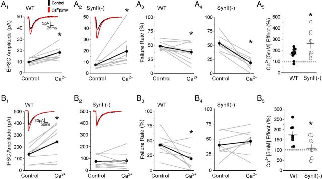
Figure 4.
Raising extracellular Ca2+ concentration mimics the effect of 4-AP application on excitatory and inhibitory transmission at WT and SynII(−) synapses. A, EPSCs recorded in slices derived from young presymptomatic SynII(−) mice exhibit an enhanced sensitivity to elevated extracellular calcium concentration (5 mm). A1, Effect of raising extracellular calcium on the amplitude of EPSCs in WT slices (p = 0.005, n = 10). A2, Effect of raising extracellular calcium on the amplitude of EPSCs in SynII(−) slices (p = 0.031, n = 8). A3, Effect of raising extracellular calcium on the EPSCs failure rate in WT slices (p = 0.047, n = 10). A4, Effect of raising extracellular calcium on the EPSCs failure rate in SynII(−) slices (p < 0.001, n = 8). A5, The effect of Ca2+ elevation is significantly stronger at SynII(−) synapses (p < 0.04). Data collected from 5 WT mice and 4 SynII(−) mice. B, Increasing extracellular Ca2+ concentration produces a robust increase in evoked inhibitory transmission in WT synapses but no detectable increase at SynII(−) synapses in slices derived from young mice. B1, Effect of raising extracellular calcium on the amplitude of IPSCs in WT slices (p = 0.033, n = 8). B2, Effect of raising extracellular calcium on the amplitude of IPSCs in SynII(−) slices (p = 0.45, n = 7). B3, Effect of raising extracellular calcium on the IPSCs failure rate in WT slices (p = 0.028, n = 8). B4, Effect of raising extracellular calcium on the IPSCs failure rate in SynII(−) slices (p = 0.5, n = 7). B5, The effect of calcium is significantly stronger at WT synapses (p = 0.026). Data collected from 5 WT mice and 4 SynII(−) mice.

