Figure 2.

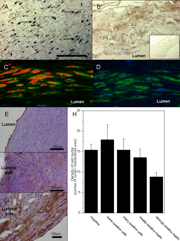
Accumulation of LDL and oxidized lipids in the aneurysm wall. Intracellular staining pattern for ApoB-100, the core protein of LDL, suggests uptake of cholesterol by cells in the aneurysm wall (A). Staining for ApoB-100 is also observed in the aneurysm wall matrix (B, negative control in the insert). Double stainings (C) for hydroxynonenal (red) and alfa-smooth muscle actin (green) demonstrate accumulation of oxidized lipids in the smooth muscle cells of the aneurysm wall (D, negative control with only alfa-smooth muscle actin in green and nuclei in blue). The accumulation of oxidized LDL (as detected by immunostaining with an antibody against copper oxidized LDL, brown) associates with degeneration of the aneurysm wall (microphotographs: E-G, association with loss of mural cells H). The bar graph H displays mean values for density of nuclei (number of nuclei per a standardized surface area) and error bars present SEM.
