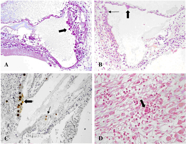
Figure 1.
CMV infection in the cochlea. A) Numerous cytomegalic inclusions in the marginal layer of the stria vascularis (arrow). Haematoxylin and eosin (HE). B) Cytomegalic inclusions in the marginal layer of the stria vascularis (small arrow) and in the Reissner’s membrane (big arrow). HE. C) CMV immunohistochemistry showing strong nuclear CMV positivity in the marginal cell layer (big arrow) and in the Organ of Corti (small arrow). In the Organ of Corti, CMV-positive cells are most likely one ciliated cell (top) and one supporting cell (bottom). The other CMV-positive cell on the right is probably a supporting cell. D) Spiral ganglion: a cytomegalic neuron surrounded by lymphocytes. HE.

