Figure 2.
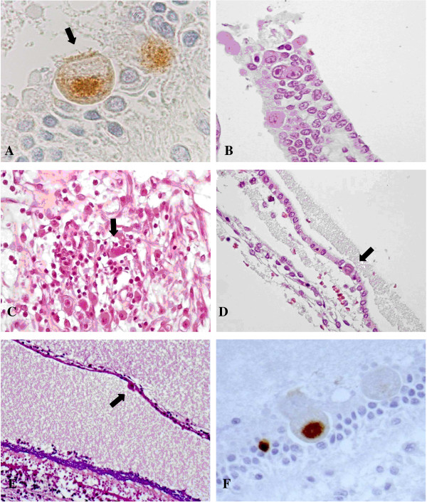
CMV infection in the vestibular apparatus. A) CMV immunohistochemistry showing CMV-positive cells within the utricle. The cell indicated by the arrow may likely be a sensory cell as suggested by the presence of cilia. B) Crista ampullaris: cytomegalic cells in the superficial epithelial layer. HE. C) Vestibular ganglion: a cluster of lymphocytes surrounding infected neurons (arrow). HE. D) One cytomegalic cell (arrow) in the epithelial layer of a semicircular canal. HE. E) Saccule: cytomegalic inclusion (arrow) in the membranous layer. On the bottom, there is the sensory macula with the otolith layer, strongly basophilic, and the hair cells underneath. HE. F) Saccule: cytomegalic cells within the macula. The positive cells may be a supporting cell (left) and a sensory cell (right). CMV immunohistochemistry.

