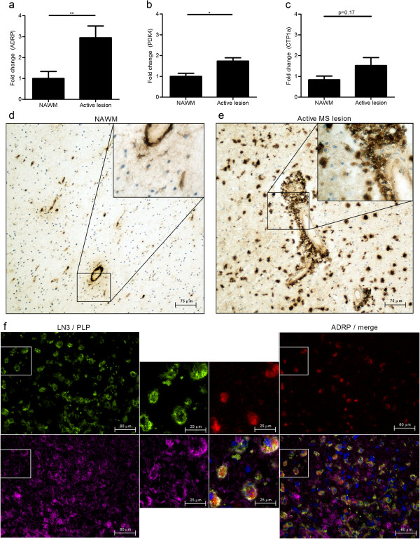
Figure 6.
PPARs are activated in myelin-phagocytosing macrophages during active MS. (a-c) Comparison of fold changes between non-demented controls (n=5) and active MS lesions (n=5). Relative quantification of ADRP (a), PDK4 (b) and CTP1a (c) was accomplished by using the comparative Ct method. Data were normalized to the most stable reference genes, determined by Genorm (YHWAZ and Rpl13a). (d,e) Normal-appearing white matter (d) and an active MS lesion (e) stained for ADRP. One representative image is shown (20× magnification). (f) Active MS lesion stained with HLA-DR (green top; left corner), ADRP (red; top right corner), PLP (magenta; bottom left corner) and DAPI (blue, bottom right corner). One representative image is shown (40× magnification).

