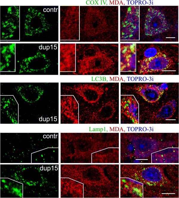
Figure 2.
MDA immunoreactivity in large pyramidal neurons in layers 3 or 5 of frontal cortex in 10-year-old individual with dup(15)/autism [dup(15)] and in 8-year-old control subject (contr) brains. Mitochondria, visualized by immunostaining for cytochrome c oxidase COX IV, were the site of weak reactivity for MDA in control but a strong reactivity in dup(15)/autism brain. Most autophagic vacuoles (detected with antibody LC3B) and lysosomes/late endosomes (detected by the presence of Lamp-1 glycoprotein) did not contain a significant fraction of MDA. Bars 10 μm.

