
Figure 2.
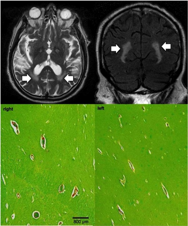
Prominent perivascular spaces evident as radial linear hyperintesities on T2 with additional perivascular confluent WMH in bilateral temporo-occipital WM (A axial T2, B coronal FLAIR). The corresponding histopathology confirms the presence of prominent perivascular spaces, yet there is no significant demyelination around the perivascular spaces, which would correspond to the confluent hyperintense T2/FLAIR signal alteration. Scale bar = 800 micrometers.