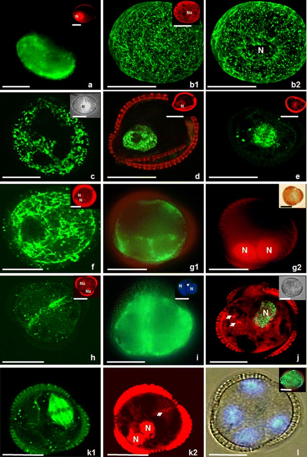
Fig. 1.

Microspores of Brassica napus induced to sporophytic development by prolonged heat stress treatment. b, e, f, h, k show fluorescent MTs; inserts in c and i, j, l show fluorescent DNA as a result of BrdU incorporation or labelling with DAPI. (a) Uni-nucleate microspore on the first day of culture. Mitochondria and ER, stained with DiOC6 (green), are around the nucleus and in the vicinity of the exine. Insert on the right hand shows the nucleus (N) after PI labelling (red). (b1, b2, c) Type II microspore with its nucleus in the cell centre. Insert in b1 on the right hand shows the nucleolus (Nu) after PI labelling (red). (b1) Dense network of thick and long bundles of CMTs in parallel orientation close to the exine. (b2) Multiple EMTs closely associated with the nucleus (N). The same microspore as in b1. (c) Microspore with a big vacuole transected by cytoplasmic strands passing from the cell centre to the cortical cytoplasm. Mitochondria and ER, stained with DiOC6 (green), are around the nucleus and in the vicinity of the exine. Insert on the right hand shows microspore with nucleus (N) in the cell centre. Microspore visualized in differential interference contrast (DIC)/UV light. (d) Type I microspore with its nucleus close to the exine. Microspore with green fluorescent nucleus indicating that it has entered S-phase. DNA synthesis is traced by BrdU incorporation and immunocytochemical labelling with Alexa 488 (green). Insert shows the nucleus (N) after PI labelling (red). (e) Dividing microspore with green fluorescent spindle MTs. Note that the orientation of the division plane turned 90° with respect to non-induced microspores. Insert shows the chromosomes in the equatorial plane after PI labelling. (f, g). 2-Nucleate endosporic structures derived from type I microspores on the second day of culture. (f) Structure with a network of densely packed cortical MTs without preferential orientation. Insert shows two symmetric nuclei (N) located close to exine after PI staining (red). (g1) Structure with DiOC6-stained mitochondria and ER in the vicinity of nuclei and in the peripheral cytoplasm. (g2) Structure with symmetric nuclei (N) after PI staining. Insert on the right hand shows the structure in DIC. (h, i) Two-celled type II structure. (h) Structure with long MTs oriented parallel to each other and aligning the plasma membrane in the equatorial plane where the new cell wall will be formed. Insert shows the symmetric position of two spherical nuclei with well visible nucleoli (Nu) after PI staining (red). (i) Structure with mitochondria and ER after DiOC6 staining in the peripheral cytoplasm of both cells. Insert on the right hand shows symmetric nuclei (N). An arrow indicates faint fluorescence of cellulose cell wall after Calcofluor White staining. (j) Two-nucleate endosporic structure in which one nucleus entered S-phase. Visualization of DNA synthesis (in green) by BrdU incorporation and immunocytochemical labelling with Alexa 488. The second nucleus is not stained and located below; only the nucleoli are visible (arrows). Insert on the right hand shows the structure in DIC. (k1, k2) Three-celled structure with one dividing nucleus within the exine. (k1) Spindle MTs are only observed at the right hand side of the structure and demonstrates asynchronous division. (k2) The same structure as on k1 after PI staining. Note chromosomes (arrow) and nuclei (N) simultaneously. (l) 4-Nucleate structure still enclosed in the exine visualized in DIC/UV on the fourth day of culture. Insert on the right hand shows the mitochondria and ER after DiOC6 staining in the vicinity of nuclei and in the peripheral cytoplasm close to the sporoderm. Bar = 20 μm
