
Fig. 6.
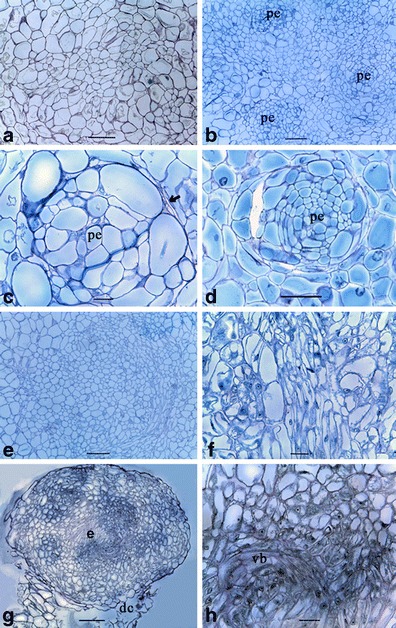
Histological observations of embryogenesis in anther-derived calli of cassava (M. esculenta Crantz). a Undifferentiated friable callus with the parenchyma cells. b The isolation of cell clusters from the other parenchyma cells giving rise to proembryos (pe). c An isolated proembryo under the higher magnification. Note the margin (arrow) that demarcates the proembryo. d The differentiation occurred in the peripheral cells of the proembryo converting them to elongated shape. e The differentiation of cells in the proembryo was continued with the cellular arrangement of the developing proembryo under higher magnification. f The degrading parenchyma cells (dc) losing contact with the developing embryo (e). g Formation of the vascular bundles (vb). Bars are as follows: a, b, e, g = 150 μm; c, f, h = 30 μm; and d = 100 μm