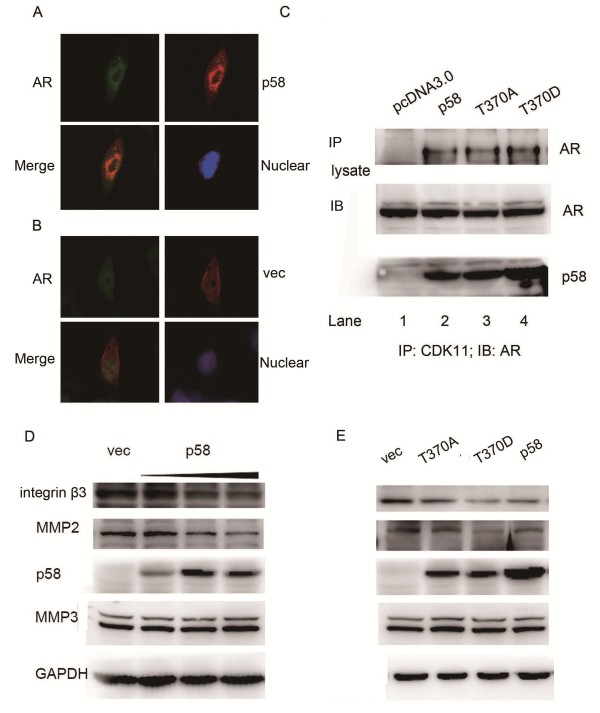
Figure 4.
CDK11p58 inhibited the expression of integrin β3 and mmp2. (A, B) LNCap cells were transfected with CDK11p58. After 48 hrs, cells were subjected to immunoflurorescent staining assay. Cells were fixed and reacted with a mouse monoclonal anti-AR antibody and a rabbit polyclonal anti-CDK11 antibody. The secondary antibodies were anti-rabbit IgG-conjugated to fluorescein isothiocyanate and anti-mouse IgG-conjugated to rhodamine red. The images were captured with a Leica confocal microscope and software provided by Leica. (C) LNCap cells were transfected with CDK11p58 and its mutants. 48 hrs later, cells were lysed and subjected to immunoprecipitation with an anti-CDK11 antibody, followed by Western-blot analysis with an anti-AR antibody in the top panel. The bottom panels showed the expression levels of the AR and CDK11p58 from the prostate cancer tissue lysates. (D) LNCap cells were transfected with pcDNA3 and CDK11p58 with increased doses. After 48 h, cells were harvested and lysates subjected to immunoblotting analysis as indicated. Protein levels were normalized to GAPDH. (E) LNCap cells were transfected with wild type CDK11p58 or its mutants. After 48 h, cells were harvested and lysates subjected to immunoblotting analysis as indicated. Protein levels were normalized to GAPDH.

