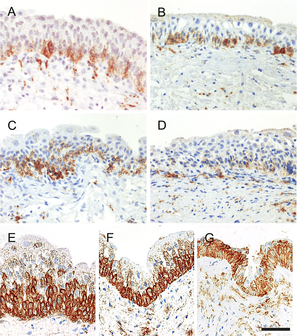
Figure 5.
Representative images of NGFR immunohistochemistry showing urothelial localisation in a range of bladder conditions including “A” non-diseased bladder taken during radical prostatectomy, “B” idiopathic detrusor overactivity and “C” stress urinary incontinence and “D” interstitial cystitis. Supra-basal expansion of NGFR labelling was only occasionally observed in idiopathic detrusor overactivity and stress urinary incontinence (see Table 2). In ketamine cystitis biopsies (“E”, “F” & “G”), supra-basal expansion of the intense NGFR labelling was observed in 10 of the 16 patients who retained intermediate urothelial cells. Scale bar in panel “G” represents 100 μm.

