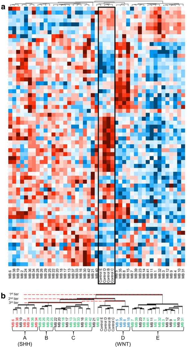
Figure 1.
GPCR expression patterns delineate distinct groups of medulloblastoma tumors. The heat map represents GPCR expression levels in 41 medulloblastoma tumors compared to normal pediatric cerebella (a). Red color indicates low CT on qPCR, corresponding to high RNA expression; blue is high CT and low RNA expression. Control cerebella (Controls B, C, D, E) are outlined in the black box. This heat map displays the results of unsupervised hierarchical clustering performed by DataAssist (v3.0; Applied Biosystems) software. Presented in this heat map are those GPCRs that were over- or under-expressed, as compared to control cerebellum, at a p ≤ 0.10 level. An enlarged image of the clustering stems, as well as the resultant tumor groups is seen in (b). Distances between tumor samples were calculated for clustering based on the ΔCT values using Pearson’s Correlation; complete linkage was used as the clustering method. Groups (A-E) of medulloblastoma tumors have emerged based solely on their GPCR expression patterns (b). Subsequent immunohistochemical subtyping of the medulloblastoma samples identified tumors of the SHH, WNT and Non-WNT/SHH subgroups (Figure 2, Table 2). The SHH- and WNT-subgroup tumors clustered tightly together on the GPCR expression heat map. Red color indicates tumors identified as belonging to the SHH subgroup, blue indicates WNT subgroup tumors and green indicates tumors identified as Non-WNT/SHH. FFPE slides not available for classification for those medulloblastoma samples in black.

