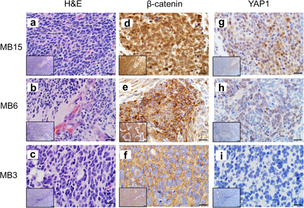
Figure 2.
Examples of immunohistochemical (IHC) staining in human medulloblastoma tumors. A method for differentiation of molecular medulloblastoma subgroups using immunoreactivity to four markers was establish by Ellison DW et al. in 2011 [32]. The H&E, β-catenin and YAP1 staining of three representative medulloblastoma tumor samples is shown in panels a – i. MB 15 (a) displays classical morphology, MB 6 (b) is an example of desmoplastic morphology and includes extensive focal nodularity, and MB 3 (c) displays anaplastic characteristics. MB 15 demonstrated nuclear and cytoplasmic immunoreactivity to β-catenin (d) and was positive for YAP1 staining (g), characterizing it as belonging to the WNT molecular subgroup. Conversely, MB 6 displayed only cytoplasmic β-catenin staining (e), and was positive for YAP1 (h), indicating that it is of the SHH subgroup. Lastly, MB 3 is cytoplasmically immunoreactive for β-catenin (f) and negative for YAP1 (i) staining, thus placing this tumor in the Non-WNT/SHH category. Staining for the above markers has been evaluated in 30/41 medulloblastoma tumors. The scale bars in panels a - i represent 20 μm; the scale bar in the inset images in each panel represents 100 μm.

