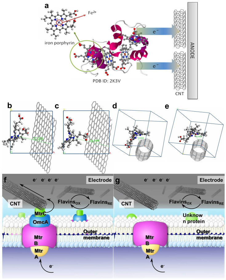
Figure 5.
Stereoview of Shewanella's small tetraheme cytochrome using the same representation as depicted in Leys et al38.: the cytochrome backbone is shown in violet, the porphyrin rings in gray, and the iron atoms in orange (a). cytochromes-graphite (Fe2+) a = 17.0668 Å, b = 17.0700 Å, c = 17.0636 Å, α = β = γ = 90 (b); cytochromes-graphite (Fe3+) a = 17.0668 Å, b = 17.0700 Å, c = 17.0636 Å, α = β = γ = 90° (c); cytochromes-CNT (Fe2+) a = b = c = 17.1123 Å, α = β = γ = 90° (d); and cytochromes-CNT (Fe3+) a = b = c = 17.1123 Å, α = β = γ = 90° (e). Schematic illustration of electron transfer at the interface of CNTs and the outer membrane of the wild-type S. oneidensis MR-1 (f); and the outer membrane of ΔOmcA/MtrC mutant (g).

