
Figure 3. Expression of VCAM-1 and binding of leukocyte-endothelial cells.
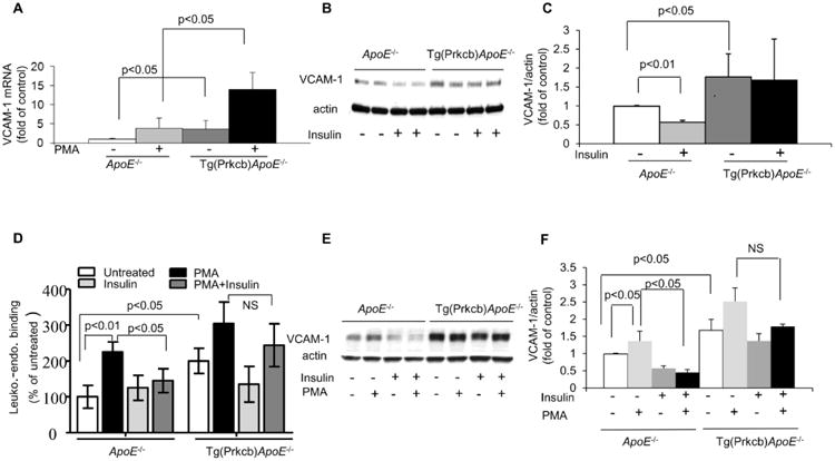
A, VCAM-1 mRNA expression in cultured lung endothelial cells. Lung endothelial cells were treated with or without 100nM PMA. VCAM-1 mRNA was determined by real time PCR and normalized by 36B4 (ApoE-/- n=8, ApoE-/- +PMA, n=3, Tg(Prkcb)ApoE-/- n=9, Tg(Prkcb)ApoE-/- + PMA n=3). (B-C), VCAM-1 protein expression in cultured lung endothelial cells. Lung endothelial cells were treated with or without 100nM insulin. VCAM-1 protein was determined by western blotting and normalized by actin. B indicates representative western blots and C shows mean value of the ratio of VCAM-1 to actin (n=4 for each group). D, Leukocyte-endothelial binding. The binding of RAW 264.7 macrophages to monolayer lung endothelial cells from ApoE-/- or Tg(Prkcb)ApoE-/- mice was measured (n=3 for each group). (E-F), VCAM-1 protein expression in endothelial cells. Endothelial cells were treated with or without 100nM insulin or 100nM PMA. VCAM-1 protein was determined by western blotting and normalized by actin. E indicates representative western blots and F shows mean value of the ratio of VCAM-1 to actin (n=3 for each group).