
Figure 5. En face Sudan IV staining of aorta.
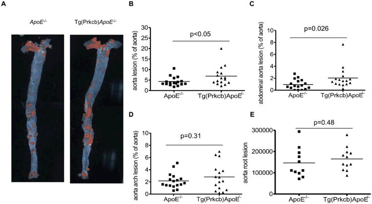
A, En face Sudan IV staining of aorta was performed in ApoE-/- (n=17) and Tg(Prkcb)ApoE-/- (n=16) mice. B, Quantitative analysis of atherosclerotic lesions in aorta (B), abdominal aorta (C), aorta arch (E) and aorta root (F) of ApoE-/- and Tg(Prkcb)ApoE-/- mice after 12 weeks of high fat feeding. Line shows the medium of the values.