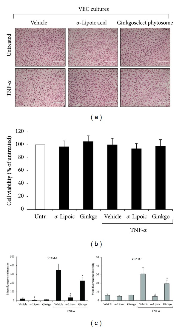
Figure 2.

Lack of cell toxicity and anti-inflammatory properties of α-Lipoic acid and Ginkgoselect phytosome in CVD-VEC model. Endothelial cells were exposed to TNF-α (5 ng/mL), Ginkgoselect phytosome, and α-Lipoic acid (both at 100 μg/mL) used either alone or in combination (1 hour of pretreatment with Ginkgoselect phytosome and α-Lipoic acid before addition of TNF-α) and analyzed after 24 hours of treatments. In (a), representative fields of cultures treated as indicated were observed by light microscopy after haematoxylin and eosin staining. In (b), cell viability is shown as percentage of untreated cultures (Untr.) set to 100%. Data are reported as means ± SD of three independent experiments. In (c), data are expressed as mean fluorescence intensity (MFI) after subtraction of background fluorescence from vehicle-treated cells. Results are reported as means ± SD of four independent experiments. *P < 0.05 compared to vehicle and TNF-α treated VEC; § P < 0.05 compared to vehicle treated VEC.
