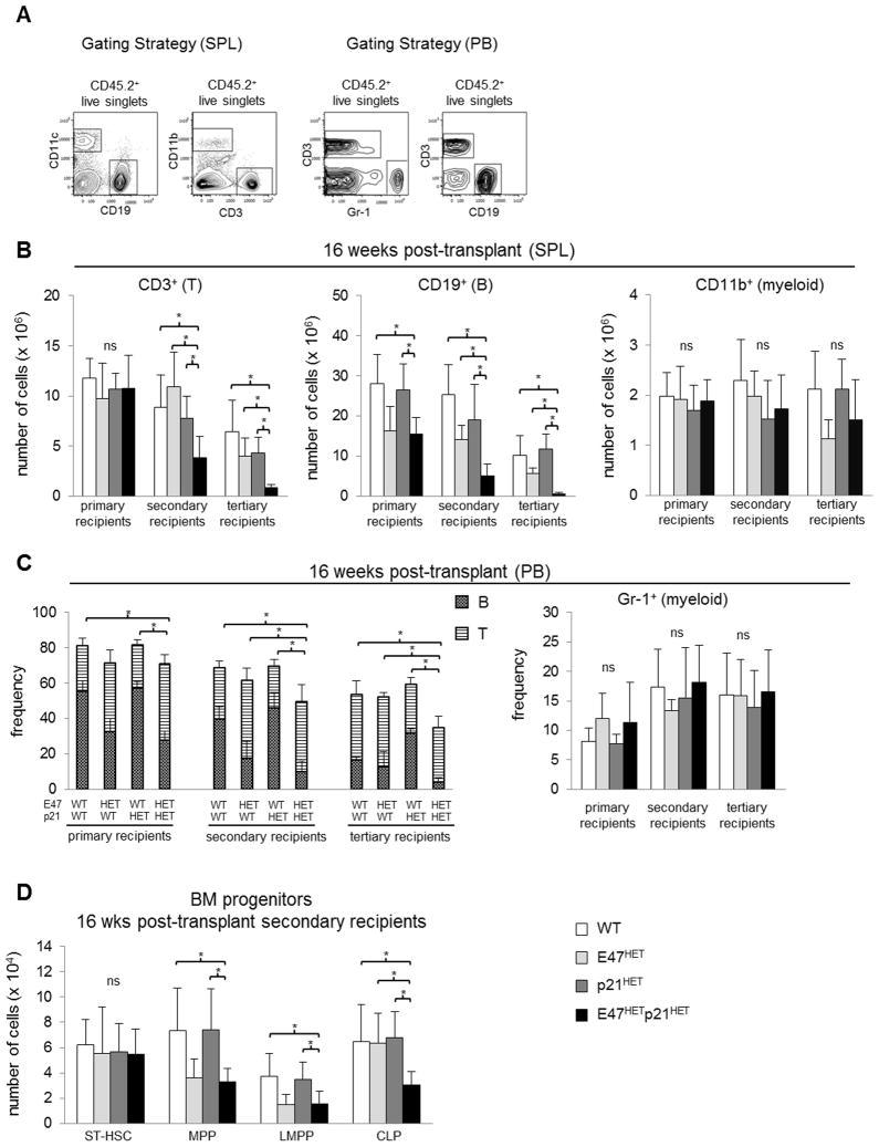
Figure 5. E47hetp21het LT-HSCs display progressive in vivo decrease in lymphoid lineage reconstitution accompanied by normal myeloid lineage reconstitution.
Serial transplantation was performed in Figure 3. Sixteen weeks after transplantation donor-derived cells were identified from spleen or peripheral blood (PB). (A) Left panel, gating strategy used to identify donor-derived B (CD45.2+ CD19+), T (CD45.2+ CD3+) and myeloid lineage (CD45.2+CD11b+) cells in spleen. Right panel, gating strategy used to identify donor-derived B (CD45.2+ CD19+), T (CD45.2+ CD3+) and myeloid lineage (CD45.2+ Gr-1+) cells in PB. Donor-derived reconstitution of lymphoid and myeloid lineage was examined in (B) spleen and (C) peripheral blood in BM recipients sixteen weeks after each round of transplantation. Graphs are shown as mean ± SD of data from n= 2–9 mice per recipient for each donor genotype. (D) Sixteen weeks after transplant, presence of donor-derived ST-HSC, MPP, LMPP and CLP in BM of secondary recipients was examined. Data are shown as mean ± SD from n= 5–9 mice per donor genotype. *p<0.05

