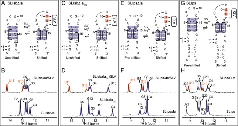
Figure 3.
NMR evidence of structural rearrangements in SLI variants. Upper panels: Proposed secondary structures of the free and SLV-bound forms of shiftable SLI [(A) SLIsbΔIa and (C) SLIsbΔIaGU] and preshifted SLI [(E) SLIpsΔIa and (G) SLIps] variants. Lower panels: Imino region of 1D 1H NMR spectra of (B) SLIsbΔIa, (D) SLIsbΔIaGU, (F) SLIpsΔIa, and (H) SLIps (bottom spectra), along with the 1D 1H 15N-filtered or 15N-edited NMR spectra of the (B) SLIsbΔIa/15N-SLV, (D) 15N-SLIsbΔIaGU/SLV, (F) 15N-SLIpsΔIa/SLV, and (H) SLIps/15N-SLV complexes for detection of SLI imino proton signals only (top spectra). The shaded imino proton signals of residues from the kissing-loop interaction (orange) and the adjacent SLI stem (purple) provide evidence for the base pairs shaded with the corresponding colors in the proposed secondary structures shown in (A), (C), (E), and (G).

