
Figure 4.
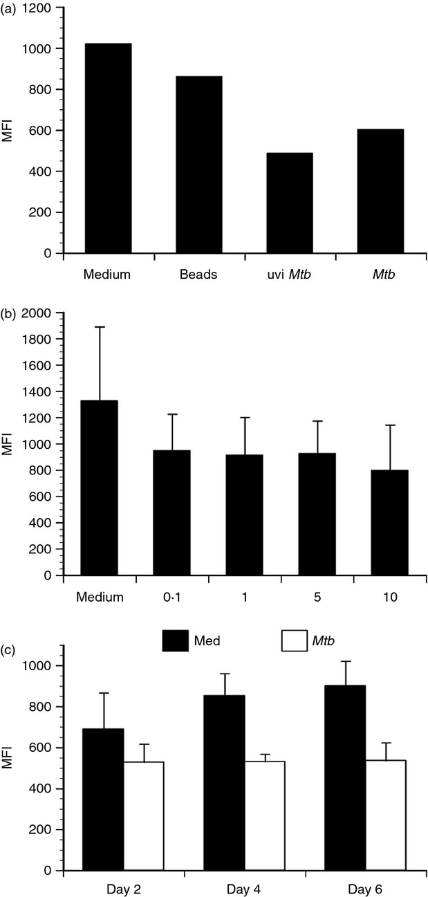
CD18 expression decrease is mediated by M. tuberculosis-derived antigen and maintained over time. Primary human dendritic cells (DCs) were treated as indicated and CD18 cell surface expression evaluated by flow cytometry. (a) A representative experiment of three performed with different donors displaying the mean fluorescence intensity (MFI) of CD18 in response to fluorescent beads, ultraviolet-inactivated M. tuberculosis (uviMtb), and live M. tuberculosis (Mtb) at a multiplicity of infection (MOI) ∼ 2. (b) CD18 MFI ± standard error in response to increasing numbers of M. tuberculosis (MOI ∼ 0·1–10) for three combined experiments with separate donors. (c) CD18 MFI ± standard error for DCs cultured in in medium alone or infected with M. tuberculosis (MOI ∼ 2) over 6 days. The data represent three combined experiments performed with different donors.