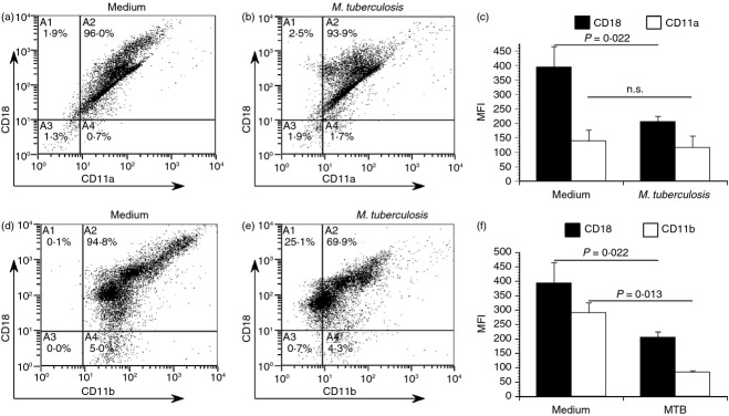
Figure 6.

Mycobacterium tuberculosis infected primary blood dendritic cells (DCs) exhibit reduced cell surface expression of both LFA-1 and Mac-1. To confirm our previous findings on blood DC, these cells were isolated from peripheral blood mononuclear cells by immunomagnetic selection. Blood DC were then either left in cell culture medium or infected with M. tuberculosis at a multiplicity of infection ∼ 2 for 48 hr. Cells were then immunolabelled for either LFA-1 (CD18/CD11a, a–c) or Mac-1 (CD18/CD11b, d–f) and analysed by flow cytometry. Scatter plots of a representative donor are shown, and the mean fluorescence intensity (MFI) ± standard error for two donors. Statistical significance in the 95% confidence interval was determined by a Student's t-test.
