
Figure 4.
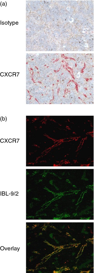
Analysis of CXCR7 protein in mouse spleen by conventional immunohistochemistry (IHC). (a) SCID mouse spleen sections were stained by conventional IHC methods with the CXCR7 monoclonal antibody (mAb) 11G8 or an isotype control mAb. 11G8 stained vessels in the red pulp. Red colour: 11G8 staining. Blue colour: nucleus counterstaining. Original magnification 200×. (b) SCID mouse spleen sections were co-stained with 11G8 and IBL-9/2, an antibody specific for splenic sinusoidal endothelial cells. An overlay of each antibody's staining pattern confirms that 11G8 stained sinusoidal endothelial cells. Original magnification 400×.