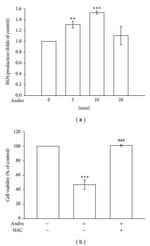
Figure 2.

The role of ROS in andrographolide-reduced cell viability in rat VSMCs. (a) Rat VSMCs were treated with 50 μM andrographolide for the indicated periods. Cells were harvested, and the formation of ROS was examined using flow cytometric analysis of DCF-DA-stained cells, as described in Section 2. (b) Cells were pretreated with a vehicle or 1 mM NAC for 30 min before being treated with 50 μM andrographolide for 48 h; cell viability was subsequently determined using an MTT assay. The results shown are representative of 4 independent experiments. The data are presented as the mean ± SEM (error bars: **P < 0.01 and ***P < 0.001, compared with the control group, and ### P < 0.001, compared with the group treated only with andrographolide).
