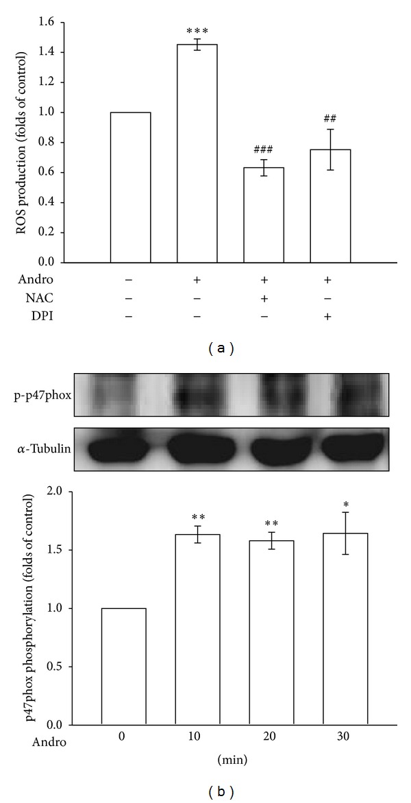
Figure 3.

Nox-mediated redox signaling in andrographolide-induced ROS formation. (a) Cells were pretreated with a vehicle, 1 mM NAC, or 10 μM DPI for 30 min before being treated with 50 μM andrographolide for 10 min, and the production of ROS was examined using flow cytometric analysis of DCF-DA-stained cells, as described in Section 2. (b) Cells were treated with 50 μM andrographolide for the indicated periods. Cells were harvested, and the phosphorylation of p47phox was examined using immunoblotting. The data are presented as the mean ± SEM (error bars: *P < 0.05, **P < 0.01, and ***P < 0.001, compared with the control group, and ## P < 0.01 and ### P < 0.001, compared with the group treated only with andrographolide).
