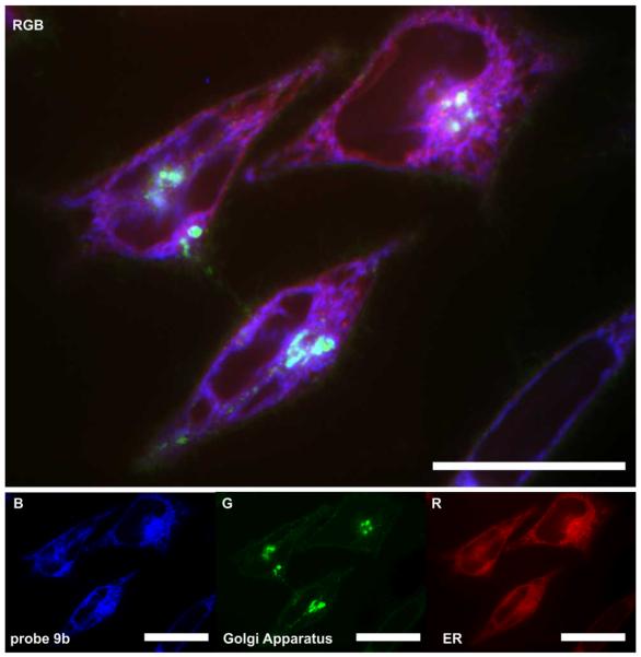
Figure 3.
Confocal fluorescent image depicting the localization of probe 12 in the endoplasmic reticulum (ER) of HeLa cells. HeLa cells were incubated with 20 nM probe 12 for 24 h at which point the cells were imaged live. Probe 12 was blue (B) fluorescent. Cells were counterstained for their ER (red, R) and Golgi apparatus (green, G) using ER-tracker Red27 and BODIPY-FL C5-ceramide28, respectively. The three-colour (RGB) image depicts the overlay of each stain. Bars denote 10 μm.

