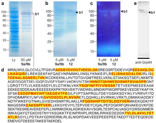
Figure 5.
Immunoprecipitation (IP) of hGrp94. (a) A Coomassie G-250 blue silver stained SDS PAGE gel depicting cell lysate from HCT-116 cells that were cultured for 24 h in media containing 50 μM probe 12. Lanes denote lysate (Ly) and the immunoprecipitated product (50 μM 9a/9b). Band b1 denotes the major protein. (b) A Coomassie G-250 blue silver stained SDS PAGE gel depicting the immunoprecipitated product obtained when using 5 μM probes 9a/9b or 5 μM probe 12. (c) A fluorescent image of the gel in (b) depicting band b1. The band at the bottom of the gel corresponds to 9a/9b or 12. (d) Trypsin-digestion followed LC-MS/MS analysis identified 11 peptides associated with hGrp94. Observed peptides are coloured and shaded. (e) While from the same protein family, the observed peptides would not be derived from cytosolic Hsp90 alpha member 1. (f) Western blot identifying the major band from the immunoprecitation with probe 12 as hGrp94. This blot depicts a Western blot using an anti-hGrp94 mAb.

