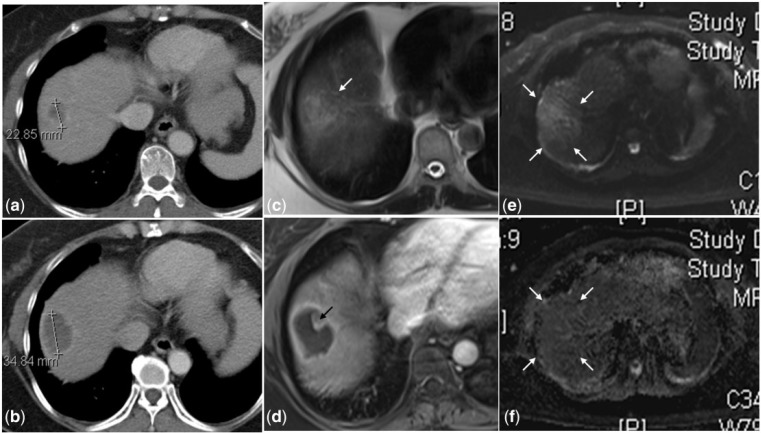
Figure 6.
A hypodense metastatic lesion is seen at the dome of the right lobe in the pre-treatment CT scan (6a) that is larger in size and continues to be hypodense at 1 month after TARE (6b). Due to the inherent hypovascularity of the tumor, it is difficult to ascertain if this represents tumor progression or necrosis. On MRI, the lesion is heterogeneous and hyperintense on the T2-weighted image (6c) with only rim enhancement on the post-gadolinium T1-weighted image (6d) suggestive of internal necrosis. The superior soft tissue resolution of MRI also depicts an eccentric solid nodule (black arrow in 6d) along the margin of this necrotic lesion that is not visible on the CT scan (6b). This indeterminate nodule shows no diffusion restriction on the diffused-weighted (6e) and ADC (6f) images, confirming the absence of any residual disease.

