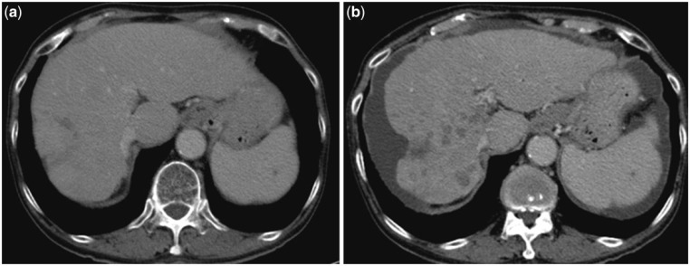
Figure 8.
Pre-treatment CT scan (8a) in a patient with multiple hepatic metastases shows normal shape and contour of the liver. CT scan obtained 2 months after bilobar 90Y radioembolization (8b) shows generalized hepatic atrophy with irregular surface. An area of prominent capsular surface retraction is noted at the dome of the right lobe. Note the several non-enhancing, ill-defined hypodense areas in the right lobe, representing areas of scarring or small bilomas. A sliver of perihepatic fluid is seen. Despite these morphological changes, this patient had normal synthetic function of the liver.

