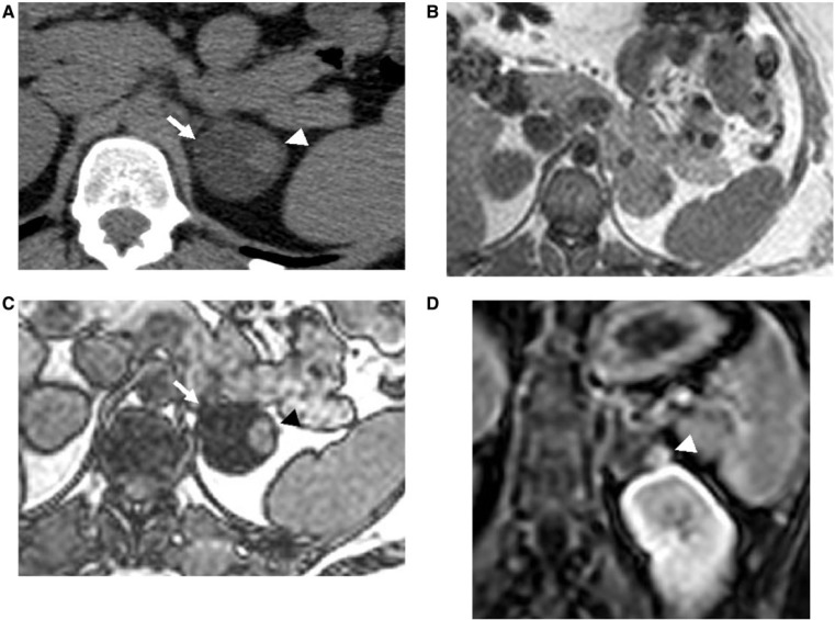
Figure 5.
A 76-year-old man with left ACT consisting of adenoma and metastases from lung carcinoma. (A) Unenhanced CT image shows a hypodense left adrenal mass (arrow) with a hyperdense nodule in the periphery (arrowhead). (B, C) Axial T1-weighted in-phase (B) and opposed-phase MR (C) images show signal drop-out of hypodense component on opposed-phase image consistent with adenoma (arrow), while hyperdense focus remains hyperintense (black arrow). (D) Gadolinium-enhanced T1-weighted fat-saturated image demonstrates enhancement of eccentric hyperdense component (white arrowhead), consistent with metastatic focus. CT-guided biopsy from different tumor components demonstrated separate adenomatous and metastatic cells on pathologic examination.

