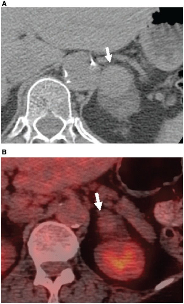
Figure 8.

Hemorrhage complicating left adrenal adenoma in a 56-year-old man. (A) Unenhanced CT image shows a hyperattenuating focus in the left adrenal adenoma (arrow). (B) FDG-PET/CT image demonstrates no increased activity within this hyperdensity, consistent with hemorrhage (arrow).
