
Fig. 3.
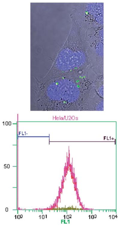
(Top) Bright-field confocal laser scanning microscopy z-scan image with fluorescent overlay of HeLa cells after incubation with PMSN (green) and with nuclei stained with DAPI (blue). (Bottom) Flow cytometry shows an internalization of the fluorescent PMSN of 98%. Fl1+ shows the fluorescent-labeled PMSN internalized cells, Fl1− shows the non-fluorescent cells.