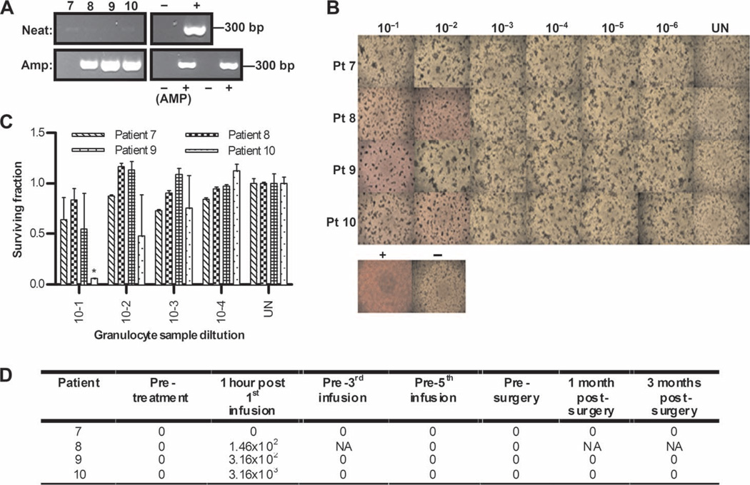
Fig. 4.
Granulocytes similarly carry replication-competent reovirus after infusion. (A) Day 1 post-infusion granulocytes were assessed for reovirus RNA by RT-PCR, using both neat and amplified samples as for PBMCs in Fig. 3A. (B) Granulocytes were assessed for functional reovirus in a TCID50 assay as for PBMCs in Fig. 3B. Photomicrographs show day 1 post-infusion granulocyte dilutions; rounded up cells and unused (red) media signify CPE. (C) Reovirus-induced cell killing by day 1 posttreatment granulocytes was further confirmed by MTT analysis. *P < 0.05 versus untreated control; error bars represent SEM. (D) Viral titers (TCID50/ml) from granulocytes over time (NA denotes samples unavailable for analysis).

