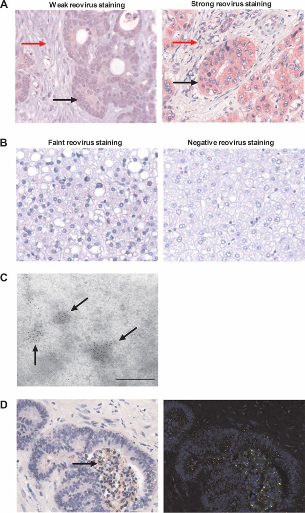
Fig. 6.
Intravenous reovirus is preferentially detected in metastatic colorectal tumor cells within the liver. (A) Immunohistochemistry images showing expression of reovirus protein (red stain) in resected colorectal liver metastases (magnification, ×400). One representative case each of weak (left; patient 1) and strong (right; patient 6) staining is shown. Malignant cells and tumor stroma are marked by black and red arrows, respectively. (B) Immunohistochemistry images for expression of reovirus protein (red stain) in normal liver (magnification, ×400). One representative case each of faint (left; patient 8) and negative (right; patient 2) staining is shown. (C) Representative EM image (patient 8) showing immunogold staining of reovirus σ3 capsid protein within colorectal liver metastases. Scale bar, 500 nm. (D) RGB image analyses of resected colorectal liver metastases, using the Nuance System (magnification, ×400). Images are representative (patient 9) and show reovirus staining (red) and caspase-3 staining (brown) (left image; arrow indicates changes of nuclear and cytoplasmic degeneration in reovirus-infected tumor cells). Right image shows conversion of RGB image to fluorescent green (caspase-3), fluorescent red (reovirus), and yellow (coexpression).

