Fig. 7.
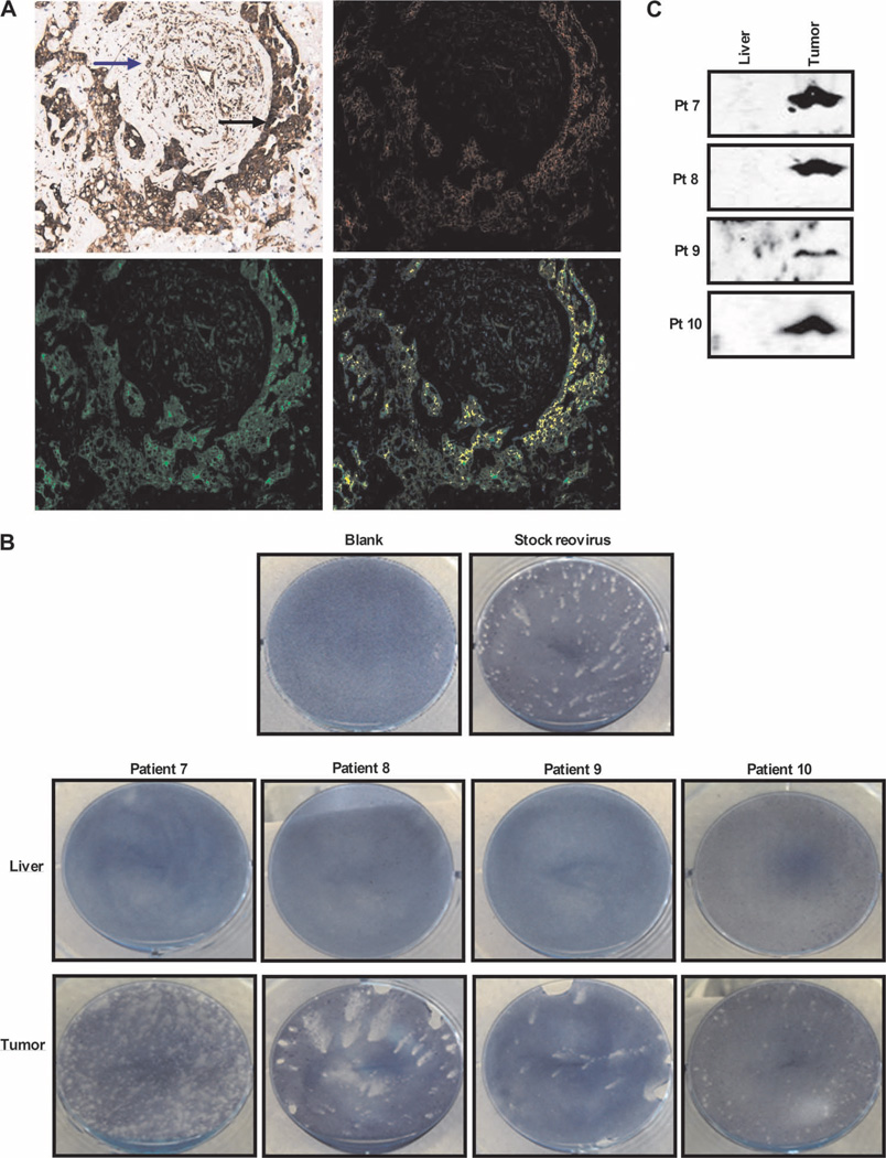
Replication-competent reovirus can be retrieved from tumor tissue. (A) RGB image analyses of resected colorectal liver metastases, using the Nuance System (magnification, ×200). Images are representative (patient 10) and show (top left) reovirus staining (red) and tubulin staining (brown) (malignant cells and tumor stroma are marked by black and blue arrows, respectively), (top right) conversion of RGB image to fluorescent red (reovirus), (bottom left) conversion of RGB image to fluorescent green (tubulin), and (bottom right) coexpression of reovirus and tubulin (yellow). (B) Plaque assays demonstrating retrieval of reovirus from freshly resected tumor and liver tissue; photographs show representative wells of optimized supernatant dilutions of 1:2500 (patient 7), neat supernatant (patients 8 and 9), and 1:1200 (patient 10). Photographs of all liver samples show representative wells of neat supernatant. (C) Culture supernatants from plaque assays performed in (B) were assessed for reovirus σ3 capsid protein by Western blotting.

