
Fig. 6.
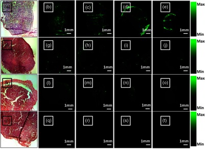
H&E and ex vivo fluorescence images of cryosections from tumors harvested at 400-min postinjection. (b–e) Images of four cryosections of the harvested tumor from mouse injected with 5 nmol ICG/mouse of targeted scVEGF-Lip/ICG. (g–j) Images of four cryosections of the harvested tumor from mouse injected with 5 nmol ICG/mouse of nontargeted Lip/ICG. (l–o) Images of four cryosections of the harvested tumor from mouse injected with 5 nmol ICG/mouse of inactive-Lip/ICG. (q–t) Images of four cryosections of the harvested tumor from mouse injected with 5 nmol ICG/mouse of free ICG. (a, f, k, p) H&E images corresponding to the first slice of each group.