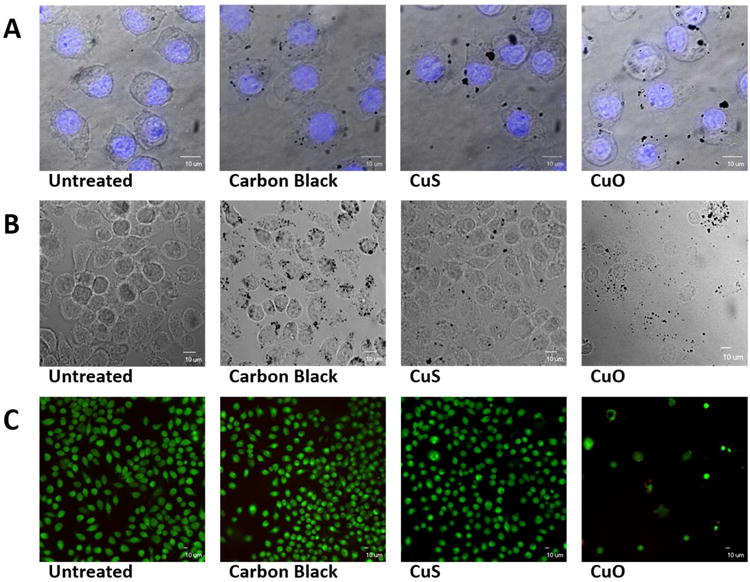
Figure 8.

Target cell uptake and toxicity of carbon black, CuS, and CuO NPs. (A) Confocal images of murine macrophages after exposure to 5 ppm of test particles for 3 hours; nuclei were visualized (blue fluorescence) using 4′6-diamidino-2-phenylindole (DAPI). (B) Brightfield microscopic images of murine macrophages 24 hours after exposure to 5 ppm of test nanoparticles. (C) Viability of target cells 24 hours after exposure to 5 ppm of test nanoparticles. Viable cells show green cytoplasmic fluorescence (Syto 10/ethidium homodimer assay).
