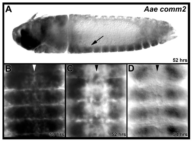
Fig. 3. Aae comm2 is expressed in the developing nerve cord.
Aae comm2 expression is detected during A. aegypti embryonic ventral nerve cord development. (A) An arrow marks nerve cord expression in a lateral view of a 52 hour old embryo oriented anterior left and ventral down. Transcripts are also detected in the brain and overlying head tissue. (B–D) Transcript levels peak in the ventral nerve cord at 50 hours (B) and are observed in a more restricted set of neurons by 52 hours (C). At 54 hours (D), when most commissural axons have crossed the midline, expression in the nerve cord has diminished. Filleted nerve cords oriented anterior upward are shown in B–D, in which the midline is marked by arrowheads.

